
Sample Limbic System Research Paper. Browse other research paper examples and check the list of research paper topics for more inspiration. If you need a research paper written according to all the academic standards, you can always turn to our experienced writers for help. This is how your paper can get an A! Feel free to contact our research paper writing service for professional assistance. We offer high-quality assignments for reasonable rates.
1. Introduction
In this research paper current ideas about the anatomical structures and functions of the limbic system will be described. The information will be limited to the limbic system as found in most mammals with greatest emphasis placed on structures prominent in the human brain. From the start it should be noted that there is less than universal agreement both about the structures that should be included in the limbic system and also about the roles played by them in mental functions and behavior. Some of the reasons for the differences of opinion among scientists arise from the many different approaches used to investigate the limbic system in both clinical and research settings. The simplest description of the limbic system is a set of highly interconnected anatomic areas lying below the cortical surface but above the most central regions of the forebrain, namely the thalamus, the hypothalamus, and areas of the ventral forebrain called the basal ganglia. Suggestions for other approaches to understanding the limbic system will be presented.
Academic Writing, Editing, Proofreading, And Problem Solving Services
Get 10% OFF with 24START discount code
2. History Of The Term, Limbic System
2.1 Early History
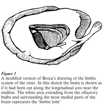
In the middle and later parts of the 1800s anatomists became interested in a region of tissue that surrounds the most medial parts of the brain, i.e., the thalamus, hypothalamus (together, the diencephalon), and the basal ganglia (caudate, putamen, nucleus accumbens, and associated regions). This medial band of tissue lies underneath the neocortical mantle and largely is separated from it by various sulci (fissures or indentations). In 1878 the French scientist Paul Broca described this deep band of tissue as a separate ‘lobe’ of the brain containing phylogenetically older cellular structures as well as the tracts that interconnected its various regions. Because it lies below the overlying neocortex it is hidden from external viewing. Broca argued that this hidden lobe was a common denominator of all mammalian brains and gave it the name ‘the great limbic lobe.’ The word limbic, which means border or fringe, was used because it is a fringe region around the deeper, more medial brain regions. Over time, many cortical regions, nuclear groups, and tracts have come to be considered components of the limbic lobe. Figure 1 shows a simplified diagram of the limbic lobe in the otter as drawn by Broca in 1878. The white area in this drawing represents the limbic lobe. From this illustration the great size of the limbic lobe is apparent, as is its strong connection with the large olfactory tracts in this animal. While in most mammals the limbic system is connected with the olfactory apparatus, the strength of the connections varies from species to species. Nevertheless the limbic lobe was originally thought to be that part of the brain associated with the sense of smell. As a result, it became known as the ‘smell brain’ or the rhinencephalon. Since the human has a relatively small olfactory system and smell was considered to be of little importance to people, it was relatively neglected by anatomists for many years.
2.2 More Recent History
It was in the late 1930s that some scientists began to think of the limbic system as being involved with emotional experiences, learning, and memory. Comparative anatomists were finding that the limbic system and its basic connections with other brain regions were quite similar in all vertebrates. This was most obvious in mammals. The great American anatomist C. Judson Herrick demonstrated similarities in the connections of limbic structures with other brain regions from the salamander through mammals (see Herrick 1956). In the salamander the most medial portions of the brain contain areas that are the primitive versions of at least three of the major cell groupings of the limbic system in all species, namely the septal area, the hippocampus, and the amygdala. Moreover, he was able to demonstrate that all of these areas had strong connections to the hypothalamus and the medial forebrain bundle.
2.3 The Medial Forebrain Bundle
In the salamander Herrick (1948) demonstrated a medial forebrain bundle that runs through the body of the hypothalamus. This bundle is found in all mammals and probably all vertebrates. It contains fibers running in both directions from the olfactory apparatus to brain stem nuclear groups, interchanging fibers with many nuclei of the hypothalamus along the way. These connections remain extensive throughout all mammalian brains. The medial forebrain bundle contains a number of powerful pathways connecting one end of the forebrain to the hindbrain. These pathways allow the forebrain and limbic system to modulate both somatic and visceral Activity and provide routes whereby the internal organs and bodily actions can influence the Activity of higher brain functions. Through the medial forebrain bundle and through other neural routes information from every sensory system reaches the limbic system.
2.4 The Papez Circuit
Research into the functions of the limbic system was greatly stimulated by a paper by Papez (1937). In it he argued that a particular set of limbic structures comprised the neural mechanism for the expression of the emotions. The pathways involved were extensive but the final projection leading to the subjective experience of emotion led from the hypothalamus to the anterior nucleus of the thalamus and then to the cingulate cortex. MacLean further championed the involvement of the limbic system with emotional behavior in a series of articles in the 1940s and 1950s (e.g., MacLean 1949, 1958). Both Papez and MacLean emphasized the importance of understanding the roles played by the cortical components of the limbic system.
3. Cortical Limbic Regions
In neuroanatomy the term cortex simply refers to tissue with neurons arranged in a layered pattern. In the human the neocortex is a thin sheet of layered neural tissue on the surface of the cerebral hemispheres. In animals with a large number of fissures about two-thirds of the total neocortex is buried within the walls of fissures. In the main, the cells of the neocortex are organized into six fairly recognizable layers. A considerable amount of cortical tissue in the forebrain is not neocortical but rather of a simpler design and with different patterns of cellular organization. Much of this non-neocortical tissue surrounds the limbic system, as seen by the white area in Fig. 1. The cingulate cortex (derived from the Latin word for belt or girdle) is one example of this more primitive cortex. It seems reasonable to consider the primitive cortical areas surrounding almost all of the limbic system structures as ‘transitional cortex’ in the sense that it comes between limbic structures and this tissue acts, in many if not all cases, as routes of entry and exit of neuronal information for the limbic structures. Other transitional cortices often mentioned are the entorhinal cortex, the periamygdaloid cortex, the pyriform cortex, and the subgenual cortex, to name but a few.
4. Nuclear Groups And Major Structures
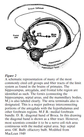
The major nuclear structures usually considered as cornerstones of the limbic system are the septal area, the amygdala, the hypothalamus, the anterior nuclear group of the thalamus, and the hippocampus. The last region is actually a primitive forebrain cortical mass (archicortex). These nuclei and some of the neural pathways between them are shown in Fig. 2. They represent what many people think of as the limbic system. But, based on both the degree of anatomical interconnections and functional considerations, many other nuclear groups have to be included to represent a reasonably complete limbic system in terms of knowledge at the start of the twenty-first century. For example, the nucleus accumbens and the ventral and medial portions of the caudate nucleus share strong neural interconnections with several major limbic regions. Functionally, they have attributes associated with limbic activities. One such attribute is their participation in the generation of feelings of pleasure.
4.1 Fiber Tracts
The major tracts of the limbic system include the fornix (hypothalamus–septalarea–hippocampus), the stria terminalis (amygdala–hypothalamus), the mammillothalamic tract (mammillary bodies of the hypothalamus to the anterior nuclei of the thalamus), and the cingulum bundle (running through the cingulate cortex from one end to the other). Although this last tract is sometimes thought to be a private association pathway for the limbic lobe, actually it carries fibers of neocortical origin forward and backward through the brain.
Of course, as described above, these nuclei, cortical areas, and tracts represent but a rough approximation of the true complexity of the regions and pathways. All of these limbic categories are far more complex than have been described above. The interested reader is referred to Paxinos (1995) for the anatomic details of the rat brain and to Crosby et al. (1962) for the human.
5. Ever-Increasing Complexity
5.1 The Consequences Of Increased Knowledge
In general, the complexity of the limbic system arises from the ever-increasing subdivisions of the larger limbic units and pathways. As new methods of identifying subdivisions of larger regions became available through advances in anatomical, physiological, and behavioral methods, finer differentiations of limbic structures were necessary. For example, as many as 22 distinct nuclear divisions within the main body of the amygdala have been proposed. But anatomical differentiation does not imply functional specificity or homogeneity within the subdivisions of anatomical areas. Even the most important, or at least best known, tracts within the limbic lobe have multiple components. For example, the large group of fibers connecting the septal area and the hippocampus has at least three separate components, that is, a large bundle just below the corpus callosum, a fiber bundle that travels for long distances in the corpus callosum, and a fiber bundle traveling over the top of the corpus callosum (the fornix longus).
5.2 Contacts With Areas Beyond Traditional Limbic Regions
As more and more has been learned about the connections of the various structures of the limbic system, it has become impossible to think of any nuclear group as an isolated entity. For example, the amygdala has strong connections with the prefrontal cortical areas, both the lateral and orbital regions, and with various neocortical and transitional cortical and neocortical regions of the temporal lobe. No unit of the limbic system operates on its own but rather in close collaboration with these other regions of the forebrain mantle. Every component of the limbic system has multiple associations with areas and nuclei below the forebrain regions, including dopamine-containing cell groups of the ventral tegmentum and the substantia nigra. The fibers from these nuclei pass through the medial forebrain bundle to the hypothalamus and the anterior extremes of the forebrain, as well as the nucleus accumbens.
5.3 Extensions Of Limbic Structures
Several years ago, the concept of an ‘extended amygdala’ was proposed (Alheid et al. 1995). The reason for the creation of this concept was that many areas, pathways, and nuclei associated with these pathways seem closely tied to specific functions of certain regions of the amygdala. They seemed to be regions that were true extensions of systems originating in the body of the amygdala, forming a unified system. Some of these associated regions were not located close to the amygdala itself. The same sort of approach could be used with the hippocampus. Some distant regions of the brain can produce similar effects to those obtained from stimulation or lesions of the hippocampus. They mimic one or more of the functions of the hippocampus. Such areas are strongly connected with the hippocampus through known neuronal pathways despite distant locations. One example would be the hindbrain sites involved in food intake and blood sugar regulation (Ritter et al. 2000). Thus, there is a basis for the concept of an ‘extended hippocampus.’ The process of creating ‘extended’ limbic areas, or for that matter ‘extended’ visual or auditory systems, would soon make every part of the brain an extension of many, perhaps all, of what are thought to be the major functional groupings of the brain. The use of ‘extended’ systems in the brain may not turn out to be a useful approach. It should not be forgotten that at all times many parts of the brain are working together to accomplish specific goals.
6. A Different Approach
The problem faced by scientists studying the limbic system is that while most have a general idea of the history and the traditional functions associated with a ‘visceral’ and emotion-related set of neural structures, there are no firm boundaries between limbic and nonlimbic forebrain regions. Evidence can be cited to implicate every area of the brain, including the neocortical mantle, in visceral, autonomic, and emotional behavior and experience. Given this situation, it is appropriate to reject an all-or-none, black or white, approach to the limbic system. It is time to recognize that every area of the brain may have involvement with ‘limbic functions’ at certain times or in certain situations.
This can be accomplished by adopting a Fuzzy Logic approach (Klir and Folger 1988) to the limbic system in which every area of the brain has some degree of limbic involvement (see Isaacson 1993). The question then becomes not whether an area is or is not a part of the system but to what degree the area is associated with limbic functions. The amount of this association may, of course, change under different circumstances. The cortical regions as well as the nuclei and pathways found in Figs. 1 and 2 would certainly have high degrees of ‘limbic-ness’ but this would not exclude their participation in other types of brain functions since they would also have varying degrees of associations with other brain systems. At the same time it would not exclude other brain areas from participating in limbic functions to some degree and at certain times. This approach would also fit with the fundamental principle of the human brain, namely that under all but the most unusual conditions, the brain’s systems operate together as a harmonious whole.
Bibliography:
- Alheid G F, de Olmos J S, Beltramino C A 1995 Amygdala and extended amygdala. In: Paxinos G (ed.) The Rat Nervous System, 2nd edn. Academic Press, San Diego, CA, pp. 495–578
- Broca P 1878 Anatomie comparee des circonvolutions cerebrales. Le grand lobe limbique et la scissure limbique dans la serie des mammifers. Review of Anthropology 1(Ser.2): 385– 498
- Crosby E C, Humphrey T, Lauer E W 1962 Correlative Anatomy of the Nervous System. Macmillan, New York
- Herrick C J 1948 The Brain of the Tiger Salamander, Ambystoma tigrinum. University of Chicago Press, Chicago
- Herrick C J 1956 The Evolution of Human Nature. University of Texas Press, Austin, TX
- Isaacson R L 1993 A ‘fuzzy set’ perspective of the limbic system: implications for cognition and memory. Neuroscience Research Communication 13(suppl.): S15–S18 (2nd edn. Academic Press, San Diego, CA, pp. 495–578)
- Klir G J, Folger T A 1988 Fuzzy Sets, Uncertainty, and In- formation. Prentice Hall, Englewood Cliffs, NJ
- MacLean P D 1949 Psychosomatic disease and the ‘visceral brain.’ Recent developments bearing on the Papez theory of emotion. Psychosomatic Medicine 11: 338–53
- MacLean P D 1958 Contrasting functions of limbic and neocortical systems of the brain and their relevance to psychophysiological aspects of medicine. American Journal of Medicine 25: 611–26
- Papez J W 1937 A proposed mechanism of emotion. Archives of Neurology and Psychiatry 38: 725–43
- Paxinos G (ed.) The Rat Nervous System, 2nd edn. Academic Press, San Diego, CA
- Ritter S, Dinh T T, Zhang Y 2000 Localization of hindbrain glucoreceptive sites controlling food intake and blood glucose. Brain Reseach 856: 37–47
ORDER HIGH QUALITY CUSTOM PAPER

Always on-time

Plagiarism-Free

100% Confidentiality